Introduction
In vivo, cells are present in a three-dimensional extracellular matrix environment rich in type I collagen, so the three-dimensional matrix is ​​a natural tissue engineering scaffold. Hydrogels are preferred as engineering tissue scaffolds because of their similar structure and good biocompatibility to most tissue natural extracellular matrices.
Type I collagen is widely found in nature and is the main extracellular matrix component in most connective tissues, with a typical hierarchical structure. The primary structure of the polypeptide chain consists of repeating (Gly-XY) n, each of which is occupied by glycine, X is usually valine, and Y is hydroxyproline. The three alpha chains are interwoven to form a triple helix, further forming filaments and fibers. The entire structure is stabilized by intermolecular hydrogen bonds and covalent bonds as well as ionic, hydrophobic and electrostatic forces between the side chain amino acid residues.
Collagen-based materials have been used in the medical field for many years, and their advantages include good biocompatibility, non-toxicity, no antigenicity and biodegradability. Collagen scaffolds can be used as substrates for cell adhesion, proliferation and differentiation, and can also be considered as ideal tissue engineering materials by in vitro reconstruction to form microfibrous structures that mimic natural extracellular matrices.
Collagen biomaterials typically require some degree of cross-linking to stabilize the structure. Chemical crosslinking can crosslink the polypeptide chains to form a network. Commonly used crosslinking agents include glutaraldehyde, EDC, N-hydroxysuccinimide, isocyanate, tannic acid and the like. In addition, photochemical crosslinking is also a common method. Enzymes can also be used for cross-linking. For example, transglutaminase can selectively catalyze the chemical reaction of glutamate and lysine on adjacent protein fibers to form a covalent amide bond and enhance the 3D matrix. Biopolymers can also be stabilized by non-covalent cross-linking bonds, such as ionic bonds formed between the chitosan amino group and the collagen carboxyl group.
The proper pH of the collagen solution promotes the occurrence of ionic reactions between protein chains. Collagen is soluble under acidic pH conditions. Therefore, under this condition, collagen cannot form an ordered hierarchical structure. Therefore, neutralization of collagen is a method of preparing gels. Under suitable conditions, the neutralization process is accompanied by aggregation of collagen molecules and alignment to form fine fibers. During this process, the addition of chemical agents optimizes the physical, chemical, and mechanical properties of the final material, but may also affect its biological properties and cell adhesion. This experiment uses dialysis as a method of neutralizing collagen solution to reduce the amount of chemical reagent used in the preparation process.
experiment
Collagen is isolated from the tail muscles of young white rats. The tendon was scraped from the adherent tissue, rinsed with deionized water, and placed in 0.1 M acetic acid at 8 ° C for 72 h. The impurities and insoluble components are removed by centrifugation. The collagen solution is then lyophilized.
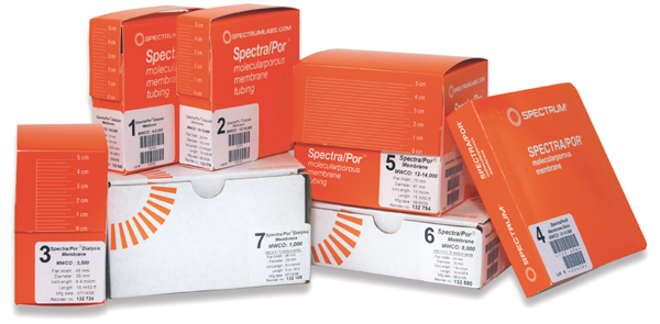
A 1% collagen - acetic acid solution (initial pH = 2.9), placed in 12-14kD Spectra / Por RC dialysis bag. To determine the optimal treatment conditions, three dialysate solutions were used: water (pH = 7), 0.1 M NaOH (pH = 13), and 10% NaCl (pH = 7), and the volume was 12 times that of the dialyzed sample. Dialysis was carried out at 6 ° C with continuous stirring and the dialysate was changed every 12 h to control the pH. The dialysis was completed after 7 days, at which time the pH of the dialysate did not change, indicating that the pH of the collagen solution was consistent with the surrounding dialysate.
Since some analytical techniques require that it be carried out under anhydrous conditions, the materials prepared are air dried or lyophilized. The collagen gel prepared by dialysis was air-dried on polyethylene sheets for 3 days (dried collagen) for FTIR-ATR, thermal analysis and contact angle detection. Three types of samples after lyophilization (freeze-dried collagen) detection: collagen with a diameter of 16 mm/length of 10 mm was used for SEM imaging, collagen with a diameter of 16 mm/1 mm was used for in vitro testing, 40 mm long/2 mm thick/5 mm wide Collagen is used for mechanical analysis.
In addition, in order to compare neutral collagen and acidic collagen, an acidic collagen film was prepared by pouring a 1% collagen-acetic acid solution onto a polyethylene sheet and air drying.
Results and discussion
There is a significant difference in the materials obtained by dialysis with different solutions: the material obtained by dialysis with 0.1 M NaOH is transparent, but semi-fluid, unable to maintain its traits; the material obtained by dialysis of 10% NaCl is white, relatively soft gel; and pure water The material obtained by dialysis is transparent, has good traits, is resistant to certain pressure, is elastic, and maintains a stable structure after incubation 24 at 37 °C. The dried neutral collagen gel forms a relatively hard, porous sponge structure. SEM imaging analysis can observe the fiber-like protrusion on the inner wall surface of the material, indicating that the collagen chain has self-organization ability during dialysis, and infrared spectroscopy also confirms that the neutral collagen structure is more ordered and contains a large number of triple helices. structure. Thermal degradation analysis of neutral collagen showed that it has a higher degradation temperature, indicating that it is more stable. The tensile strength test for neutral collagen also showed that there are multiple interactions in the internal collagen chain, which stabilizes the protein structure. Freeze-dried/dried neutral collagen has a strong coefficient of expansion. In addition, in vitro biological testing of collagen materials has shown that cells can tolerate neutral collagen.
The experimental results show that a stable and transparent collagen hydrogel can be prepared by the dialysis process, insoluble in water and PBS buffer, and can be significantly expanded. By changing the pH of the collagen solution, a more ordered structure can be obtained, which is similar to the triple helix of natural collagen, and the thermal stability of the material is better than that of the acidic collagen film. After the gel is lyophilized, a porous, steric, and shape-stable material is obtained, and the material is suitable for cell growth, but since the surface of the material is non-porous, the cells do not migrate into the internal structure of the stent.
The dialysis neutralization method can also be used to prepare hydrogels in which collagen and other proteins are mixed, such as elastin and glycosaminoglycans. These mixtures are polyelectrolytes, so ionic and electrostatic reactions can occur between molecules, changing the structure of the collagen. The addition of different macromolecules may have a positive or negative effect on the hydrogel, but should not be added too much to avoid changing the stability of the hydrogel, and the molecular weight should be large enough to prevent passage through the membrane pores during dialysis. And the loss.
The editor compiles and introduces this article for communication purposes. Due to the limited level, please be aware of any inconvenience. For details, please refer to the original text.
Original: Skopinska-Wisniewska J., Olszewski K., Bajek A., et al., Dialysis as a method of obtaining neutral collagen gels. Material Science and Engineering C, 2014, 40: 65-70.


Scan, pay attention to the official digital WeChat public account, get more application information!
Incubator Products,Air Circulation System,Lighting Incubator Machine For Lab,Incubator Temperature Controlle
Guangdong Widinlsa International Co.Ltd , https://www.guangdongwidinlsa.com